

Authored by Nicholas Oliver Corey Kuzik
Reviewed by Stephen Carrell, PGY-5 at University of Alberta
Edited and posted by Yosra Er-reguyeg
Oct 12, 2026 | 5 min

Part 6: Diplopia on Your Ophthalmology Elective
There is good news and bad news when it comes to diplopia. Good news: working through the differential diagnosis is a fairly systematic process. Bad news: that systematic process can feel like some sort of labyrinth when you are fumbling through your cosplay as a qualified orthoptist.
Thankfully, as a medical student, you do not need to solve the entire labyrinth. You need to figure out a few important things. First and foremost, is the diplopia monocular or binocular. If binocular, then you will need to go further into the differential:
And, as always, is there anything here that should make me worried?
First: Monocular or Binocular?
Monocular diplopia is double vision that remains when the affected eye is fixating by itself. This means there is an optical problem in that eye:
Monocular diplopia isn’t less complicated per se, but it does typically narrow the area of the problem down to refractive and media abnormalities in that specific eye. Check vision with pinhole to see if it improves. Apply fluorescein to check tear break-up time, and examine for corneal abrasions/scarring. Use the slit lamp to verify if they have a cataract or any irregularities in the vitreous or retina.
Binocular diplopia is double vision that disappears when either eye is covered. This indicates there is a problem with the eyes working together, or the two eyes are not properly aligned. Typically, each eye sends visual information to the brain, and the cortical fusion mechanisms join the two images together. This fusion creates the illusion of 3-D vision or stereopsis. Misalignment of the eyes can send images that are too out-of-sync for the brain to process, resulting in diplopia. Misalignment generally means the muscles involved with keeping the eyes aligned are not doing their job. Possible causes include:
If your patient presents with binocular diplopia, you now need to try to isolate where the issue is. To help isolate the issue, you should try to observe a deviation at rest, while covering one eye at a time, and in different cardinal planes of extraocular movements.
Just Act Normal
Before busting out the advanced moves, look at the patient. Is one eye obviously turned in, out, up, or down? If you’re not very good at playing spot the differences, you can use the corneal light reflex to help out. Actually, even if you’re good at playing spot the differences, you should use the corneal light reflex so you don’t look like a fool for mislabelling a deviation that is actually pseudo strabismus. Also, ask them whether the diplopia is vertical, horizontal, or diagonal. This can help isolate the area of misalignment.

Corneal Light Reflex
Have the patient look directly at a small light source. The reflection of the light should be in roughly the same location on both corneas. If the reflections are asymmetric, the eyes may be misaligned.
For example:
This is a quick screening test. It will not tell you everything, but it can confirm that you are not imagining the misalignment. But there could still be a very subtle misalignment, or one that is intermittent.

Cover it up
This is one of the major ways to isolate a tropia or a phoria. Tropia is persistent, think about when you visit the tropics and never want to leave. Phoria is intermittent, I don’t have a clever way to remember that, you’re on your own.
Cover-uncover test
Cover one eye while watching the uncovered eye. If the uncovered eye moves to pick up fixation, it was misaligned while both eyes were open. That is a tropia, or manifest strabismus. For example, if the uncovered eye moves outward to fixate, it must have been sitting inward beforehand: an esotropia. Repeat on the other side.
Alternate cover test
Now move the cover rapidly from one eye to the other, preventing the patient from regaining binocular fusion. Watch for refixation movements each time the cover switches (meaning watch the eye that you are uncovering). This can reveal a phoria, a tendency for the eye to drift when fusion is interrupted. The distinction is useful:
A longstanding phoria can eventually decompensate and cause intermittent diplopia, particularly with fatigue or age.
Extraocular Movements: Comitant or Incomitant?
Returning to our eye vitals, one should get a sense of each eye's ability to look in all directions. All binocular diplopias should have their extraocular movements tested. You do not need to become a strabismus specialist, but this distinction is useful. Check extraocular movements to see whether the misalignment worsens or remains the same across the different cardinal positions of gaze.
Comitant
The amount of misalignment is roughly similar in different directions of gaze. Think more about:
Incomitant
The misalignment magnitude changes depending on where the patient looks. For instance, let’s say in the primary position there is a left esotropia of some magnitude. If as the patient looks right the magnitude decreases and as the patient looks left the magnitude increases. This would point to the only affected/palsied muscle being the left lateral rectus. Overall, seeing a incomitant misalignment should have you thinking more about:
If you are seeing binocular diplopia with incomitant misalignment, now is a good time to think about cranial nerve palsies.
The Big Three Cranial Nerve Palsies
There are three main cranial nerve palsies to know about the 3rd, 4th, and 6th. To help remember how to isolate these three, it’s important to know what they innervate. The 4th innervates the Superior Oblique muscle, the 6th innervates the Lateral Rectus muscle, and the 3rd innervates the Rest of the ocular muscles. It helps to remember this by thinking of it like a chemical formula: SO4-LR6-R3.
CN VI Palsy
This is probably the easiest one to recognize. CN VI innervates the lateral rectus (LR6), which abducts the eye. For instance, if the right sixth nerve is weak:
So:
Horizontal diplopia + impaired abduction = think CN VI.
Some patients compensate by turning their head toward the affected side.
CN IV Palsy
CN IV innervates the superior oblique (SO4). This one can be much subtler, since the primary ocular movement of the superior oblique is intorsion. Patients typically describe:
Unlike a sixth nerve palsy, the eyes may look almost normal in primary gaze. If the diplopia is vertical, you can compare the misalignment in right and left gaze and with right and left head tilt to help localize the affected muscle. As a medical student, recognizing:
Vertical diplopia + compensatory head tilt = consider CN IV
👁️💪
Want to flex? Bust out the three step test for identifying SO palsies: https://eyewiki.org/Three_Step_Test_for_Cyclovertical_Muscle_Palsy
CN III Palsy
CN III controls the rest (R3):
A complete third nerve palsy can therefore produce:
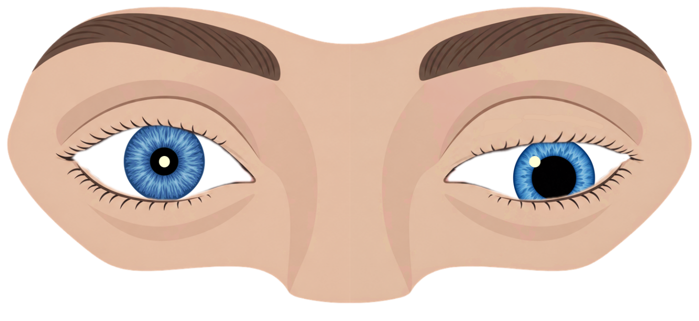
This is also the cranial nerve palsy where the pupil really matters.
A new third nerve palsy associated with a dilated or poorly reactive pupil, particularly when accompanied by headache or pain, should raise concern for a compressive lesion such as an aneurysm and needs urgent investigation.
Do not get so excited about correctly identifying the nerve that you forget to look at the pupil.
What About Myasthenia?
Myasthenia gravis can imitate almost any pattern of binocular diplopia.
Useful clues include:
One useful ophthalmology pearl:
Myasthenia does not affect the pupil.
So ptosis and diplopia that is worse at night with normal pupils should put it on your differential. You do not need to diagnose myasthenia from across the room. Just remember it when the pattern refuses to behave like a clean cranial nerve palsy.
Do Not Forget the Orbit
Not every eye that fails to move has a weak nerve. Sometimes the eye physically cannot move because something is restricting it. Think about orbital disease or entrapment when diplopia is accompanied by:
Causes include:
A restricted eye and a paretic eye can look similar at first. That is another good reason to examine the whole patient rather than just focusing on extraocular movements.
What Should Make You Worried?
Diplopia deserves more urgent assessment when there is:
On the other hand, isolated monocular diplopia from an optical cause or clearly longstanding/comitant strabismus without concerning features can usually be assessed less urgently.
Knowing what needs attention now versus what can wait is just as useful as naming the nerve.
If You Remember Nothing Else
Monocular diplopia: Double vision remains with one eye closed. Usually refractive or medium abnormalities.
Binocular diplopia: Goes away with one eye closed. The eyes are misaligned.
Corneal light reflex: Observe misalignment when light reflection differs from eye to eye.
Extra Ocular Movements: Appreciate which direction of gaze is compromised, and if it gets better/worse/remains consistent across the cardinal positions of gaze.
while moving
Cover-uncover: finds a tropia.
Alternate cover: can find a phoria.
And recognize the big three. SO4-LR6-R3:
Then ask:
Is this a routine alignment problem, or is there something here that makes it urgent?
You do not have to escape the entire orthoptics labyrinth. You just need to know who to punt it to next.
Next month: The Abnormal Pupil on Your Ophthalmology Elective


